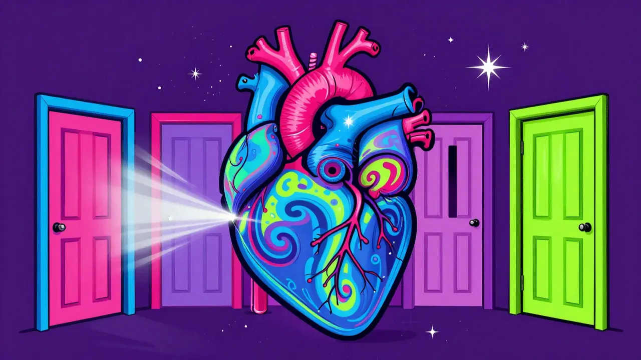
Imagine your heart is a house with four doors. For the blood to flow correctly, these doors must open wide to let traffic through and close tight to keep it from leaking back. When those doors get stuck or broken, you have heart valve disease, which is a condition where one or more of the heart's four valves malfunction due to narrowing (stenosis) or leakage (regurgitation). It’s not just a medical term; it’s a mechanical failure that forces your heart to work overtime. If left unchecked, this extra strain can lead to heart failure, irregular rhythms, or worse. The good news? We have better tools than ever to fix these "doors," ranging from tiny catheters to advanced surgical replacements.
You might be reading this because you heard a murmur during a checkup, or perhaps a loved one was diagnosed recently. You probably want to know: Is it serious? What does stenosis actually mean versus regurgitation? And if surgery is needed, what are my options? Let’s break down the mechanics, the symptoms, and the modern treatments without the confusing jargon.
Understanding the Mechanics: Stenosis vs. Regurgitation
To understand the problem, you first need to understand the parts. Your heart has four main valves: the aortic valve, mitral valve, tricuspid valve, and pulmonary valve. Each plays a specific role in keeping blood moving in one direction. When they fail, it happens in one of two ways.
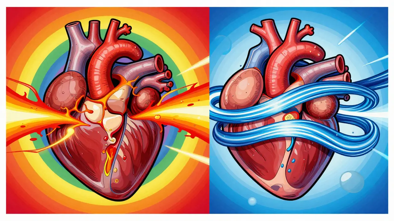
Stenosis is like a door that won’t open all the way. The leaflets become stiff, often due to calcium buildup, narrowing the opening. This restricts forward blood flow. Your heart has to pump much harder to push blood through that small gap. Over time, the muscle thickens and weakens.
Regurgitation (also called insufficiency) is like a door that doesn’t latch shut. Blood leaks backward into the chamber it just left. This causes volume overload-your heart fills up with too much blood, stretches out, and eventually loses its efficiency.
| Feature | Stenosis | Regurgitation |
|---|---|---|
| Mechanism | Narrowing of the valve opening | Incomplete closure causing leakage |
| Blood Flow Impact | Restricts forward flow (obstruction) | Causes backward flow (volume overload) |
| Primary Heart Strain | Pressure overload (muscle thickens) | Volume overload (chamber stretches) |
| Common Symptoms | Chest pain, fainting, shortness of breath | Fatigue, palpitations, swelling |
| Most Affected Valve | Aortic valve | Mitral valve |
The Big Two: Aortic and Mitral Valve Issues
While any valve can fail, the aortic valve and mitral valve are the most commonly affected. Knowing which one is involved changes the symptoms and the treatment plan significantly.
Aortic Stenosis is the most common serious form of valve disease, affecting about 2% of adults over 65. In younger patients under 70, half of these cases are caused by a bicuspid aortic valve, a congenital condition where the valve has two flaps instead of three. As you age, calcium builds up on these flaps. Severe aortic stenosis is defined medically by a valve area smaller than 1.0 cm². Without treatment, the 5-year survival rate for severe untreated aortic stenosis drops to just 50%. With timely replacement, that jumps to 85%.
Mitral Regurgitation is different. Here, blood leaks back into the left atrium when the ventricle contracts. It’s often more subtle in early stages. Patients report extreme fatigue-79% of them-because their body isn’t getting enough oxygen-rich blood forward. It can be primary (the valve itself is damaged) or functional (the valve is fine, but the heart chamber has stretched so much the valve can’t close). The COAPT trial showed that for functional cases, a transcatheter repair device called MitraClip reduced mortality by 32% compared to medication alone.
Recognizing the Warning Signs
Valve diseases are often called "silent killers" because symptoms don’t appear until significant damage has occurred. However, there are clear signals your body sends when the valves struggle.
- Shortness of Breath: This is the most common complaint. You might feel winded climbing stairs or even lying flat in bed (orthopnea).
- Fatigue: If your heart isn’t pumping efficiently, your muscles don’t get enough fuel. You’ll feel tired after minimal effort.
- Chest Pain (Angina): Common in aortic stenosis. The heart muscle needs more oxygen but can’t get it through the narrowed valve.
- Fainting (Syncope): A dangerous sign. It means your brain isn’t getting enough blood flow during exertion.
- Palpitations: Feeling like your heart is racing or fluttering. This can signal atrial fibrillation, a common companion to valve disease.
If you experience chest pain, fainting, or sudden severe shortness of breath, seek emergency care immediately. These are red flags that your heart is struggling to compensate.
Diagnosis: How Doctors Find the Problem
You likely won’t guess you have valve disease. It usually starts with a doctor hearing a heart murmur during a routine exam with a stethoscope. A murmur is the sound of turbulent blood flow. But a murmur isn’t a diagnosis-it’s a clue.
The gold standard for diagnosis is an echocardiogram. This ultrasound of the heart allows doctors to see the valves moving in real-time. They measure:
- Valve Area: How wide the opening is.
- Pressure Gradient: The difference in pressure across the valve.
- Ejection Fraction: How well the heart pumps overall.
If the echo shows severe issues, you may need a CT angiography or cardiac MRI to get a detailed map of the anatomy before planning surgery. Early detection is critical. Waiting for symptoms in aortic stenosis reduces 2-year survival rates significantly.
Surgical and Interventional Options
Treatment depends on severity, age, and overall health. Mild cases are monitored. Moderate to severe cases require intervention. The landscape of treatment has changed dramatically in the last decade.
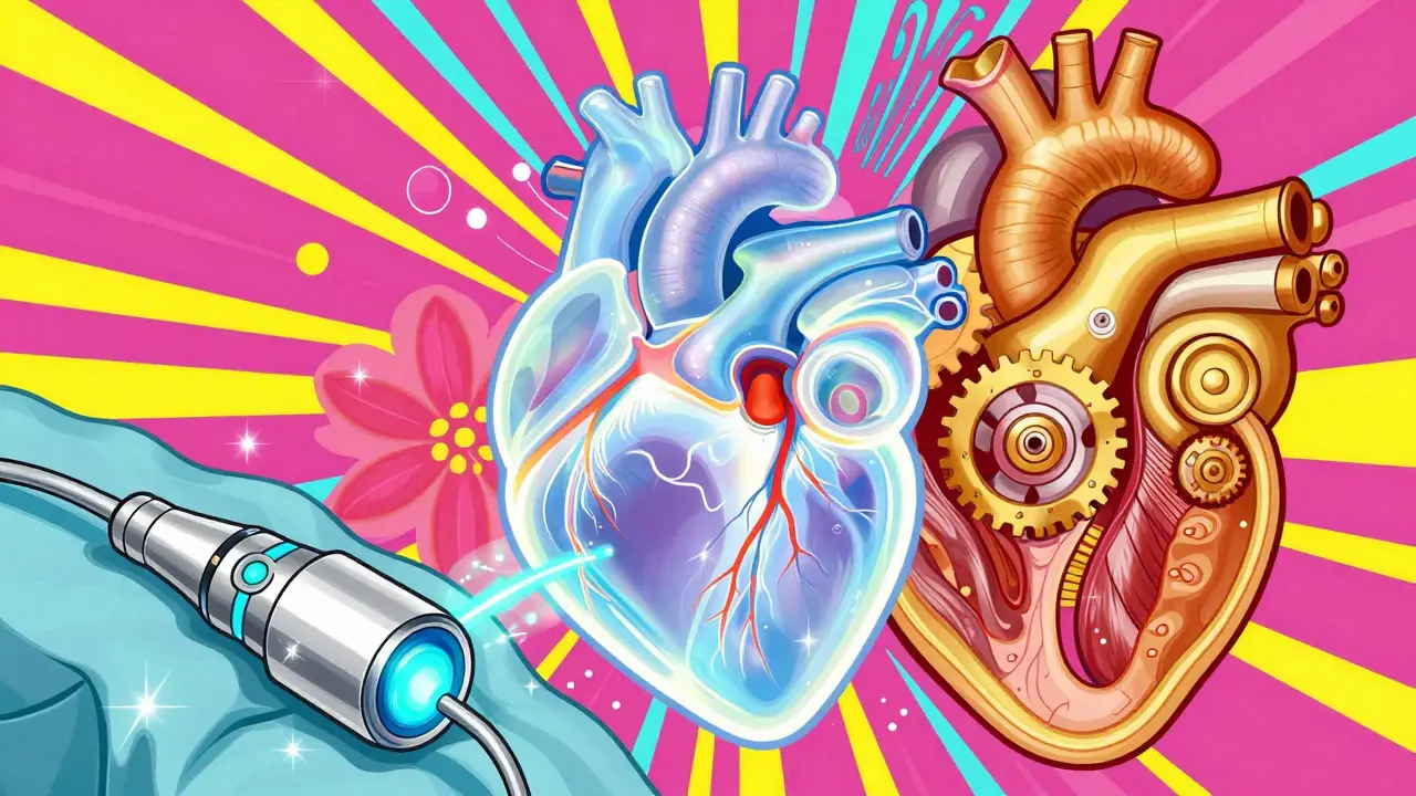
Transcatheter Aortic Valve Replacement (TAVR): This is a game-changer. Instead of open-heart surgery, doctors insert a new valve through a catheter in the groin. It’s minimally invasive, with a hospital stay of just 2-3 days. Initially reserved for high-risk elderly patients, recent trials (like PARTNER 4) show it’s now safe and effective for lower-risk patients aged 60-80. By 2030, experts predict 80% of valve procedures will be transcatheter-based.
Surgical Aortic Valve Replacement (SAVR): Traditional open-heart surgery. It requires cracking the sternum and using a heart-lung machine. Recovery takes weeks. However, for younger patients or those with complex anatomy, surgery remains the gold standard. You choose between a mechanical valve (lasts forever but requires lifelong blood thinners) or a bioprosthetic valve (made from animal tissue, lasts 10-15 years, no long-term blood thinners needed).
Valve Repair: Especially for the mitral valve, repair is preferred over replacement if possible. Techniques include annuloplasty (tightening the ring around the valve) or using clips (like MitraClip) to bring the leaflets together. Repair preserves the native valve structure and avoids the risks of prosthetic valves.
Medication: Drugs cannot fix a broken valve. They manage symptoms-reducing blood pressure, controlling heart rate, or reducing fluid buildup-but they do not stop the progression of the disease. They are a bridge to surgery, not a cure.
Life After Treatment
Recovery varies. TAVR patients often report significant energy improvements within 30 days. Surgical patients face a longer road, with sternotomy pain lasting up to 8 weeks. Post-procedure life involves regular follow-ups. If you have a mechanical valve, you’ll take warfarin and monitor your INR levels closely. If you have a bioprosthetic valve, you may only need antibiotics before dental procedures to prevent infection.
The key takeaway? Don’t ignore the signs. Valve disease is progressive. Once symptoms start, the clock is ticking. Modern medicine offers excellent solutions, but timing is everything.
What is the difference between stenosis and regurgitation?
Stenosis is the narrowing of a heart valve, which restricts blood flow forward and forces the heart to pump harder. Regurgitation is the leakage of a heart valve, allowing blood to flow backward, which causes the heart chambers to stretch and fill with excess volume.
Is TAVR surgery safer than traditional open-heart surgery?
For many patients, especially those over 75 or with higher surgical risk, TAVR (Transcatheter Aortic Valve Replacement) is safer and less invasive. It involves a small incision in the groin rather than opening the chest. Recent studies show comparable survival rates to open surgery even in lower-risk patients aged 60-80, with faster recovery times.
How long do artificial heart valves last?
Mechanical valves are durable and can last a lifetime, but they require lifelong blood-thinning medication. Bioprosthetic (tissue) valves typically last 10 to 15 years and do not require long-term blood thinners, making them a popular choice for older adults who want to avoid daily medication risks.
Can heart valve disease be cured with medication alone?
No. Medications can help manage symptoms like high blood pressure, fluid retention, or irregular heartbeats, but they cannot repair a physically damaged or calcified valve. Once valve disease becomes severe, mechanical intervention (repair or replacement) is necessary to restore proper function and improve survival rates.
What are the most common symptoms of aortic stenosis?
The classic triad of symptoms for severe aortic stenosis includes angina (chest pain), syncope (fainting or dizziness), and dyspnea (shortness of breath), particularly during physical activity. Fatigue is also very common. If you experience these, especially fainting, you should seek immediate medical evaluation.